Published by
Stanford Medicine

Author

Medical Education, Stanford News, Videos

Whiz Kids: Investigating healing mechanisms in the oral mucosa

whiz-kids-investigating-healing-mechanisms-in-the-oral-mucosa

Over the past week I’ve been showing off projects from Stanford’s Clinical Anatomy Research Scholars, a program that brought 15 interns to campus this summer to do research alongside faculty in a variety of fields across biomedicine.

In this final installment of the “Whiz Kids” series, high-school student Julia Gross does a wonderful job explaining the research she did on the healing mechanisms in the oral mucosa.

The CARS program is based in the Division of Clinical Anatomy and is directed by W. Paul Brown, DDS.

Previously: Whiz Kids: Developing a program for “young anatomists”, Whiz Kids: Research looks at handling pediatric crises effectively, Whiz Kids: Creating an iBook about the heart, Whiz Kids: Using haptics for surgical simulation and Whiz Kids: Teaching anatomy with augmented reality

Medical Education, Stanford News, Technology, Videos

Whiz Kids: Developing a program for “young anatomists”

whiz-kids-developing-a-program-for-young-anatomists

As regular readers have seen over the last week, I’ve been highlighting projects from the the Clinical Anatomy Research Scholars (CARS) program at Stanford. That program allowed 15 interns to come campus this summer to do research alongside faculty in a variety of fields across biomedicine.

Those students also produced YouTube videos on their work and entered them in NPR’s “What’s Your Big Idea?” contest. In this video, Ruby Moreno, an incoming Stanford undergraduate student, explains the program she developed to teach middle school students about anatomy.

The CARS program is based in the Division of Clinical Anatomy and is directed by W. Paul Brown, DDS.

Previously: Whiz Kids: Research looks at handling pediatric crises effectively, Whiz Kids: Creating an iBook about the heart, Whiz Kids: Using haptics for surgical simulation and Whiz Kids: Teaching anatomy with augmented reality

Medical Education, Stanford News, Videos

Whiz Kids: Research looks at handling pediatric crises effectively

whiz-kids-research-looks-at-handling-pediatric-crises-effectively

Over several previous posts, I’ve highlighted projects from the the Clinical Anatomy Research Scholars (CARS) program at Stanford. If you haven’t seen my earlier entries, CARS allowed 15 interns to come campus this summer to do research alongside faculty in a variety of fields, including medicine, surgical simulation, robots, and biomedical visualization.

Those interns also made short YouTube videos describing the work and entered them in NPR’s “What’s Your Big Idea?” contest. In this video, Colleen Hamilton explains her research project with the Center for Advanced Pediatric & Perinatal Education, which focused on how to handle pediatric crises effectively.

The CARS program is based in the Division of Clinical Anatomy and is directed by W. Paul Brown, DDS.

Previously: Whiz Kids: Creating an iBook about the heart, Whiz Kids: Using haptics for surgical simulation and Whiz Kids: Teaching anatomy with augmented reality

Medical Education, Stanford News, Videos

Whiz Kids: Creating an iBook about the heart

In earlier Scope posts, I’ve shown off projects from the the Clinical Anatomy Research Scholars (CARS) program at Stanford. CARS allowed 15 interns to come campus this summer to do research alongside faculty in a variety of fields, including medicine, surgical simulation, robots, and biomedical visualization. Those students also made their own video presentations describing the work and entered them in NPR’s “What’s Your Big Idea?” video contest.

In this video, Gunn High School student Nikki Murthy talks about the iBook she created about the heart. The book covers the vascular system, basic heart anatomy, heart diseases and more. Murthy and her collaborators also used 3D bioimaging to enrich the book. It’s an impressive feat - and it’s even more so when you realize Murthy is just 17 years old!

The CARS program is based in the Division of Clinical Anatomy and is directed by W. Paul Brown, DDS.

Previously: Whiz Kids: Using haptics for surgical simulation and Whiz Kids: Teaching anatomy with augmented reality

Medical Education, Stanford News, Videos

Whiz Kids: Using haptics for surgical simulation

whiz-kids-using-haptics-for-surgical-simulation

In an earlier post, I highlighted one very impressive augmented-reality project from the the Clinical Anatomy Research Scholars (CARS) program at Stanford. Through CARS, 15 lucky interns are invited to campus to conduct research alongside professors in a variety of fields, including medicine, surgical simulation, robots, and biomedical visualization.

Over the next week, I’ll be highlighting more video presentations from the students. In this video, high-school student Matthew Miller talks about his project studying how haptic technologies might improve surgical simulation.

This video has also been entered in NPR’s “What’s Your Big Idea?” video contest. You can get a sense of the videos submitted from NPR’s contest playlist.

The CARS program is based in the Division of Clinical Anatomy and is directed by W. Paul Brown, DDS.

Previously: Whiz Kids: Teaching anatomy with augmented reality

Medical Education, Stanford News, Technology, Videos

Whiz Kids: Teaching anatomy with augmented reality

whiz-kids-teaching-anatomy-with-augmented-reality

As part of the Clinical Anatomy Research Scholars (CARS) program at Stanford, 15 lucky interns came to campus this summer to conduct research alongside professors in a variety of fields, including medicine, surgical simulation, robots, and biomedical visualization.

This video shows off one such project by Monte Vista High School student Nishant Jain. In his project, Jain developed an augmented reality application that can be used to teach anatomy. Here’s how he describes his handiwork on YouTube:

In order to combat the challenges of current anatomical educational techniques, I have developed a prototype of an educational tool that has the ability to superimpose accurate digital models of human anatomy, that are derived from actual patient CT Scan Data, into real space, in real time. This will enable students to interact with a digital model as if it was present right in front of them, thereby providing a natural and intuitive experience that invokes spatial learning. Combined with annotations and video lectures by Stanford faculty that could be embedded in the software, this augmented reality system is a significant novel development that has the potential to revolutionize anatomical education.

As you can see, it’s pretty neat work. Jain, along with his fellow interns, also entered his project in NPR’s “What’s Your Big Idea?” video contest. I’ll be sharing more of these projects on Scope in the coming days. Right now, Jain’s video has 1,691 views and 255 likes!

The CARS program is based in the Division of Clinical Anatomy, and is directed by W. Paul Brown, DDS.

Sports

Tennis, anyone? New York Times examines tennis for the blind

There are three reasons why I love this time of year: the French Open wraps up on Sunday, followed by Wimbledon at the end of June and the U.S. Open at the end of August. Boring? Hardly! Thrilling? Frequently. The agility and strength of the athletes is jaw dropping as they slam the ball back and forth with precision and power, and sweat and grunts, delivering shots that make me marvel over and over again.

The current Grand Slam action definitely puts me in a tennis state of mind, so I was very interested in an article in the New York Times about a way to play tennis that had never occurred to me: blind tennis. Fortunately, it’s occurred to people who are blind or who have limited vision - and it’s growing in popularity.

Thomas Lin writes:

Blind tennis is made possible, scientists say, by the adaptability of the human brain - which appears to repurpose its visual area, the occipital cortex, to process sound and touch in response to blindness.

A series of studies discovered activity in the visual cortex when blind test subjects read Braille, and found that a blind woman could no longer make sense of the raised dots after suffering an occipital stroke. Another study, of sighted subjects who were blindfolded, showed that the occipital cortex began processing tactile and auditory information within five days.

If you missed the story or you haven’t checked it out, I highly recommend doing so - and the story has a nice video that’s worth watching.

Events, History, Sleep, Stanford News

An afternoon with bedheads and Deadheads

an-afternoon-with-bedheads-and-deadheads

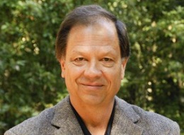
Yes, it might seem like sleep researcher William Dement, MD, PhD, and the late Jerry Garcia would make very strange bedfellows. But, that wasn’t the case at a Stanford event on Saturday. There, they blended together - albeit, in a circular way - like a sweet dream in a deep sleep.

More than 60 people with a variety of ties to the Stanford Sleep Disorders Clinic and Research Center came together at the Jerry House (yes, that Jerry) for the unveiling of a long-awaited plaque discussed earlier today. I was one of the people there to honor the Stanford “sleep camps” held there in the 1970s and ’80s.

A wide variety of those involved with the camps showed up at the event to revisit their pasts and talk about their presents. It was a fun, wacky reunion, bringing together 10 years worth of researchers and researchees. Some flew in from distant ports, and their entrances generated hugs and squeals and hearty handshakes. There was a coterie of “campers,” some the progeny of professors and staff, who happily dispensed memories and swapped tales. There were full-fledged doctors who, as undergrads, acted as “sleep counselors.” And there were sleep-research luminaries who were, back then, just getting the sleep-research field powered up.

The plaque, which was concealed under a very ’60s tie-died cloth, was unveiled, and the researchers who led the work at the camps spoke, acknowledging the importance of the research and expressing gratitude to all involved. The remarks of Mary Carskadon, PhD, expanded into a very detailed string of stories - a decade of escapades involving rambunctious kids, stealthy undergrads, and 24-hour-a-day volleyball tournaments.

There was an abundance of delicious food, a terrific Sancerre, a quantity of beer and an enormous, mega-cake with a stunning replica of the plaque laid out in the frosting. It was the perfect fuel for dancing on a sunny, spring-like afternoon, so when the Grateful Dead-inspired band let loose, people were ready. The air was charged, and the past quickly became the present. Gauging by the expressions on many faces, I don’t think I was the only one transported back to college days!

Previously: Thanks, Jerry: Honoring pioneering Stanford sleep research
Photo of Dement by Robert Tognoli

Humor, Stanford News

Pumpkin Jobs: Stanford med student’s carving tricks - a real treat!

It’s obvious why Amy Ladd, MD, professor of orthopaedic surgery, sees the potential in Raymond Tsai. The third-year medical student from Northridge, Calif., is a whiz with sharp objects. And, Halloween pumpkins. He carved his first one in 2006—a portrait of Steve Irwin, the croc hunter. Two years ago at the annual first-year student pumpkin-carving extravaganza, he blew away classmates with a carved rendition of Michael Jackson. Tsai’s latest Jack-o’-lantern masterpiece pays homage to the late Apple co-founder and creative genius Steve Jobs, pictured above in both the glowing and unlit versions.

Tsai starts his process by finding a photo of the carvee that has well-defined value contrasts. From the photo he makes a template, then heads off to scour the pumpkin patch. “I’m picky about my pumpkins,” he explains. “It has to be big enough to fit my template, and its shape needs to be oblong to fit the face. The tricky part is finding one without the deep grooves.”

For the Jobs job, Tsai spent almost two hours surveying the pumpkin’s terrain. The actual carving took about three and a half hours. “I focus on the most defining characteristics,” he says. “The beard and the straight-on gaze were the don’t-screw-up bits.” He did cut off one nostril by accident, but said it didn’t make a notable difference. As soon as the last detail is carved, the entire pumpkin gets bathed in bleach to keep it fresh. Occasional spritzes are applied PRN. “Steve Irwin got moldy really quickly, so I went online and read that bleach helps slows down decomposition.”

Now that he’s finished with his seasonal gourd carving, he’s looking forward to his upcoming rotation in internal medicine and to his duties as Speaker of the AMA Medical Student Section. He also seems like a likely candidate to join forces with the folks featured on Science Friday’s recent video pick.

Photo courtesy of Raymond Tsai

Humor

Move over, Vesalius

scope_artist_goldfish.jpg

Over the last couple of years I’ve been dazzled by the anatomical imagery that’s come out of the School of Medicine. From the pristine photos in the Bassett Collection, to the wizardry of Paul Brown, DDS’ group, the images have shown us our innards and fascinated our brains.

Now, thanks to Katherine Wells’ article in Science of the Arts, I’ve discovered a whole new galaxy of truly amazing dissections. Artist Jason Freeny is exhibiting a beautifully rendered dissection of a Carassius auratus auratus, bis coquere, otherwise know as a goldfish. What a rush! They’re so easy to eat by the handful that I admit I’ve never inspected the inside of one. Now, I can see that it’s not the cheddar that gives them their cheesy goodness; it’s their little bones and guts! Brilliant. I tell you, this guy’s a genius. Check out his website for a whole range of dissection wackiness.

Image courtesy Jason Freeny

Stanford Medicine Resources: