Yesterday’s announcement about Stanford scientists developing a process that renders tissue, specifically a mouse brain, transparent spurred a significant amount of excitement among both the scientific community and general public. We’ve captured the reactions in tweets, blog posts, videos and quotes from new articles on our Storify page.
Among the video content is an interview with Karl Deisseroth, MD, PhD, explaining the work, a fly-through of a complete mouse brain using fluorescent imaging, and commentary from Michelle Freund, PhD, a project officer in the National Institute of Mental Health Division of Neuroscience and Basic Behavioral Science, discussing the significance of the work. Mixed in with the videos are remarks from experts about how the breakthrough will advance the field of neuroscience and other research applications and candid comments from Twitter users. We hope the collection provides a broader perspective on the research and its potential to revolutionize cell biology.
Earlier today I wrote about a breakthrough method called CLARITY, pioneered by Stanford psychiatrist/bioengineer Karl Deisseroth, MD, PhD, for rendering intact tissue samples transparent. Above is a video clip showing off the new method’s capabilities. First you’ll witness a “fly-through” of a complete mouse brain using fluorescent imaging. The immediately following clip - it’s spectacular! - provides a three-dimensional view of a mouse hippocampus (the brain’s brain’s memory hub), with projecting neurons depicted in green, connecting interneurons in red, and layers of support cells, or glia, in blue.
Note that in both cases, there was no need to slice the tissue into ultra-thin sections, analyze them chemically and/or optically and then laboriously “sew” them back together via computer algorithms in order to reconstruct a 3-D virtual image of the biological sample. All that was required, after performing the necessary hocus-pocus, was to ”send in the stain” (i.e., use histochemical means to paint different cell types different colors) and move the sample or camera lens or shift the latter’s focal length. Nice trick. With big implications for biomedical research.
Stanford psychiatrist and bioengineer Karl Deisseroth, MD, PhD, spent much of this century’s first decade developing a revolutionary method for studying the brain: optogenetics. In 2010, Nature Methods heralded optogenetics as its “method of the year.”
It looks as though lightning has struck the Deisseroth lab again.
Suppose, just for a moment, that you’re conducting espionage on a heavily guarded multi-story building strongly suspected to be an advanced nuclear-weapons facility. The building quickly proves utterly inaccesible. Fortunately, you manage (through methods too covert to be revealed here) to procure a floor plan. Nice going. Now, you know a lot about the floors themselves and a bit of cross-sectional detail on the bases of whatever’s sitting on them. Better than nothing.
Now, imagine - in fantasyland, anything goes - that you can don goggles enabling you to peer right through the building’s outer walls and directly observe its three-dimensional structure, including its concealed laboratories and the instruments and manufacturing machinery inside of them. Payday!
An analogous technique developed by Deisseroth promises to revolutionize cell biology. Exploring connections among, and contents within, the billions of cells in a chunk of tissue often involves slicing the chunk into ultra-thin sections, exposing each slice’s top and bottom surfaces for microscopy or histochemical and electrical manipulation. Sophisticated computation can stitch the slices back together (virtually), roughly reconstructing the sample’s three-dimensional structure. (That’s the floor plan I mentioned earlier.)
Unfortunately, all this sawing disrupts key connections within the tissue and distorts its constitutent cells’ geography. Plus, while those sections are thin, they’re not infinitely thin. Light and chemicals can penetrate only so far. Volumes of valuable information about their innards remains concealed.
Deisseroth’s paradigm-shifting method, called CLARITY, renders tissue transparent while leaving it structurally intact, yet accessible to large “detective” molecules scientist use to gain information about cells’ surface features and genetic contents. In a study just published in Nature, a group led by Deisseroth (who discusses his work in the video above) converted an entire adult mouse brain into an optically transparent, histochemically permeable replica of itself. The position and structure of proteins embedded in the membranes of cells and their intracellular organelles remained intact.
Okay, step back with me for a minute. Essentially, all cells are liquid-filled bubbles of oil. (Nerve cells are better visualized as long, branching, liquid-filled tubes whose walls are made of fat.) These oil/fat (in science-speak, “lipid“) bubbles and walls (“membranes”) both house and compartmentalize their contents, so operations inside them can be carried out in relative isolation. Dotting membranes’ surfaces are all kinds of proteins performing innumerable activities key to the health of the cells they enclose and the tissues those cells compose.
Evolution designed lipid membranes to be mostly impermeable to large molecules, and they happen to be opaque (or else we’d all be transparent). In a feat of chemical engineering, Deisseroth’s team replaced the lipids with, for all purposes, clear plastic. With their work, you could literally read a newspaper through the mouse’s brain. Formerly membrane-bound proteins remained anchored in the membranes’ doppelgangers, retaining their structures (a big deal, as a protein’s structure determines its function). The tissue was also nanoporous: It permitted bulky “reporter”molecules such as stain-carrying antibodies and strips of DNA to flow deep into the transformed tissue sample and out again.
Obviously you wouldn’t want to try this on yourself, although Plastic Man certainly seems to have worked out the kinks.
In case you missed it, an article Technology Review offers a closer look at how scientists are taking computing beyond mechanics and electronics into the realm of living cells. Katherine Bourzac reports on efforts by Stanford biologists to develop methods for programming cells to detect diseases or make adjustments in fermentation tanks used to manufacture drugs or other chemicals.
… [R]esearchers at Stanford University have developed a way to make genetic parts that can perform the logic calculations that might someday control such activities.
The Stanford researchers’ genetic logic gate can be used to perform the full complement of digital logic tasks, and it can store information, too. It works by making changes to the cell’s genome, creating a kind of transcript of the cell’s activities that can be read out later with a DNA sequencer. The researchers call their invention a “transcriptor” for its resemblance to the transistor in electronics. “We want to make tools to put computers inside any living cell—a little bit of data storage, a way to communicate, and logic,” says Drew Endy, the bioengineering professor at Stanford who led the work.
Further down in the story Endy explains how the work could be used to improve diagnosis and treatment of cancer.
Stem-cell therapy for damaged hearts is a brilliant idea whose time has not yet come. To date, human and animal trials - there have been quite a few - in which stem cells were injected into cardiac tissue to treat severe heart attacks or substantial heart failure have mostly produced poor results.
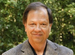
Stanford’s Sam Gambhir, PhD, MD, who heads the medical school’s Department of Radiology, thinks he knows part of the reason why, and he may have found a way around it.
At present, there’s no way to ensure against faulty initial placement, he told me in an interview about his study describing the proposed solution, just published in Science Translational Medicine. “You can use ultrasound to visualize the needle through which you deliver stem cells to the heart. But once those cells leave the needle, you’ve lost track of them.”
As a result, key questions go unanswered: Did the cells actually get to the heart wall? If they did, did they stay there, or did they diffuse away from the heart? If they got there and remained there, for how long did they stay alive? Did they replicate and develop into heart tissue?
Gammbhir’s lab has figured out a way to “mark” stem cells before infusing them into the heart, rendering them visible to standard ultrasound as they’re squeezed out of the needle. The key was to invent an innovative imaging agent, in the form of nanoparticles whose diameters clustered just below one-third of a micron — less than one-three-thousandth the width of a human hair. The nanoparticles’ main ingredient, silica, shows up on ultrasound. The particles were also doped with the rare-earth element gadolinium, so they can also be observed using MRI.
It turns out that mesenchymal stem cells — a class of cells often used in heart-regeneration research because they can differentiate into beating heart cells and because they can sometimes be harvested directly from the patients themselves, avoiding possible immune-compatibility problems — were happy to gobble up the nanoparticles in a lab dish without losing any of their ability to survive, thrive, and replicate themselves.
When Gambhir and his associates infused these nanoparticle-loaded stem cells into the hearts of healthy mice, they were indeed able to monitor the cells via ultrasound after they left the needle tip, guide them to the targeted area of the heart wall, and still get a strong MRI signal from the cells two weeks later.
Stem-cell therapy for damaged hearts isn’t going to be cheap anytime soon. (A wild guess of, say, $50,000 per procedure is probably not too far off the mark.) But a one-time delivery, if it worked, could replace a lifetime of constant drug administration. Adding what Gambhir estimates might be another $2,500 a pop for the added imaging capability is likely to be hugely cost-effective, because it could greatly improve the odds of the procedure’s success.
You’ve heard of Sputnik, that little tiny antenna-clad chunk of metal heaved into low orbit on October 4, 1957, effectively kicking off the Space Age?
Well, make way for Gutnik. A news release issued by NASA’s Ames Research Center foretells the launch into space of a satellite inhabited by a bunch of nano-mariners who, left to their own devices, would no doubt rather curl up inside a bowel.
Sometime in the next one to three years, according to the release, a so-called “nanosatellite” weighing about 30 pounds and peopled by the intestinal bug E. coliwill streak into the sky, with the mission of amassing data on whether the zero-gravity environment that cloaks our planet might increase microbes’ resistance to antibiotics. That’s important, because, as the release states:
Bacterial antibiotic resistance may pose a danger to astronauts in microgravity, where the immune response is weakened. Scientist believe that the results of this experiment could help design effective countermeasures to protect astronauts’ health during long-duration human space missions.
E. coli is probably the most-studied micro-organism in all of science. While most strains are harmless and actually quite friendly (producing vitamin K for us, just to name one of the nice things they do), some of them can cause food poisoning, urinary-tract infections and more.
Gutnik (whose real name is EcAMSat) is the brainchild of Stanford microbiologist A.C. Matin, PhD, the principal investigator for the joint NASA/Stanford University School of Medicine project. Matin’s previous inventions include microbes capable of gobbling up environmental toxins like uranium and chromium, as well as magnetic-field-seeking bacteria that can increase the contrast of magnetic-resonance imaging. So this new satellite caper is just one more in a series of wild but potentially very useful feats of imagination.
The thing that really knocks me out, though, is how all these scientists and engineers will manage to get those billions of little tiny bugs to sit still while the chin straps on their little tiny space helmets are being fastened.
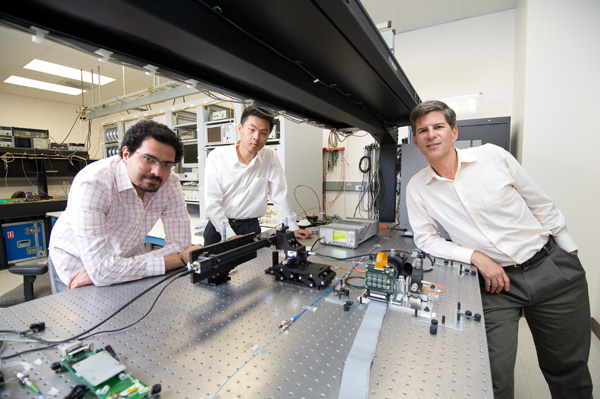
Two years ago, a discussion about biophotonics between Joseph Kahn, PhD, a Stanford professor of electrical engineering, and fellow engineer Olav Solgaard, PhD, raised the question of whether it was possible to send light through a hair-thin fiber, form a bright spot inside the human body, and record images of living tissue. The challenge surrounding the development of such a device sparked both their interest, and they began working to turn the conceptual idea into reality.
A Stanford Reportarticle published today offers a closer look at the engineers’ effort to develop a prototype of the single-fiber endoscope that boosts a resolution that is four times better than existing instruments:
The so-called micro-endoscope is a significant step forward in high-resolution, minimally invasive bio-imaging, with potential applications in research and clinical practice. Micro-endoscopy could enable new methods in diverse fields ranging from study of the brain to early cancer detection.
…
Their prototype can resolve objects about 2.5 microns in size, and a resolution of 0.3 microns is easily within reach. A micron is one thousandth of a millimeter. By comparison, today’s high-resolution endoscopes can resolve objects only to about 10 microns. The naked eye can see objects down to about 125 microns.
Kahn, Solgaard and colleagues recently published a paper (.pdf) on the prototype in the journal Optics Express.
Researchers at Massachusetts General Hospital have developed a tethered, pill-sized endoscope that creates detailed images of the esophageal wall. The imaging system, which is enclosed in a capsule about the size of a multivitamin pill, allows doctors to map a person’s esophagus in microscopic detail within a few minutes.
A paper (subscription required) published yesterday in Nature Medicine describes researchers’ work. The video above illustrates how the device works, it’s advantages over traditional endoscopy and it’s potential use for screen patients for Barrett’s esophagus, a precancerous condition usually caused by chronic exposure to stomach acid.
A new approach using a soft and squishy material called hydrogel may help rebuild healthy cartilage, according to findings reported this week in Science Translational Medicine.
An article published today in the San Jose Mercury News describes how the material works to mend damaged cartilage and keep joints moving. In the piece, Garry Gold, MD, professor of radiology at Stanford and a co-author of the new study, discusses the research and says the approach shows promise in becoming the standard for a simple, low-cost procedure. Ryder Diaz writes:
Unlike other parts of the body, cartilage does not receive a supply of blood, which carries the nutrients needed for healing.
…
One common method, called microfracture, drills tiny holes into the bone in the spot where the cartilage is missing. Blood and stem cells from the bone marrow spill into the damaged area. Surgeons hope these cells will grow into new, healthy cartilage.
But because there is nothing for the cells to hold onto, they often grow in a sloppy way, creating scar tissue. “The scar cartilage, it’s less durable,” [Jennifer Elisseeff, PhD, a professor at Johns Hopkins University and a co-author of the study] said.
That poor-quality cartilage can break down and disappear in two years, Gold said. Patients can experience the return of pain and the arrival of arthritis as more healthy cartilage disappears.
The new procedure tested by Gold, Elisseeff and colleagues added hydrogel to the microfracture technique.
Here, exposed bone is coated with a primer before holes are drilled. Then the gooey liquid hydrogel is poured in to fill in the missing cartilage, sticking to the primer. Light is used to set the gel, similar to the way a dentist uses light to solidify a filling in the cavity of a tooth.
The hydrogel creates a scaffold for the blood and stem cells from the bone marrow to cling to as they grow into healthy, new cartilage, Gold said. As new cartilage grows, the hydrogel dissolves.
After six months, the team found that patients treated with hydrogel had less pain and more healthy cartilage filled the pothole than the traditional approach.
Researchers at Stanford have created a synthetic material that is sensitive to touch and capable of healing itself after being torn or cut. A paper published yesterday in the journal Nature Nanotechnology describes how chemical engineering professor Zhenan Bao, PhD, and her team combined the self-repairing ability of a plastic polymer and the conductivity properties of a metal to develop the new material, which could lead to more advanced prosthetics.
A Stanford Reportstory discusses how Bao and her group tested their new product’s mechanical strength:
The researchers took a thin strip of the material and cut it in half with a scalpel. After gently pressing the pieces together for a few seconds, the researchers found the material gained back 75 percent of its original strength and electrical conductivity. The material was restored close to 100 percent in about 30 minutes. “Even human skin takes days to heal. So I think this is quite cool,” [Benjamin Chee-Keong Tee], a researcher on the projectsaid.
What’s more, the same sample could be cut repeatedly in the same place. After 50 cuts and repairs, a sample withstood bending and stretching just like the original.
The composite nature of the material created a new engineering challenge for the team. Bao and her co-authors found that although nickel was key to making the material strong and conductive, it also got in the way of the healing process by preventing the hydrogen bonds from reconnecting as well as they should.
For future generations of the material, Bao said, the team might adjust the size and shape of the nanoparticles, or even the chemical properties of the polymer, to get around this trade-off.