Published by
Stanford Medicine

Author

In the News, NIH, Research

NIH Director polls Twitter for real-world responses to budget cutbacks

nih-director-polls-twitter-for-real-world-responses-to-budget-cutbacks

Here’s a developing social media story of interest to scientists, clinicians and the general public. National Institutes of Health Director Francis Collins, MD, PhD, kicked a hornets’ nest on Twitter earlier today with a tweet asking researchers to describe the direct impact of the U.S. budget sequestration, which began in March, on their research and lives. He asked respondents to use the hashtag #NIHSequesterImpact. The responses (some of which I’ve included below) are fascinating and depressing:

I am no longer encouraging undergraduates to consider graduate school. No future in it.

The NIH training grant I’m on was canceled

Watching top notch science go unfunded; bright, young investigators forced to close labs, it’s heartbreaking.

I know a lot of very smart USA young researchers that are seriously considering China

Nothing will impact treating patients more in the long term than poorly funded basic science. Nothing

Check it out if you’d like to hear a real-time conversation about what it’s like to be a researcher today, and join in if you have anecdotes to share.

Previously: As budget sequester nears, a call for Congress to protect funding for scientific and medical research, Director of NIH discusses accelerating translation of biomedical research into clinical applications and Francis Collins profiled in New Yorker

Cancer, Research, Stanford News

“Housekeeping” protein complex mutated in about 1/5 of all human cancers, say Stanford researchers

housekeeping-protein-complex-mutated-in-about-15-of-all-human-cancers-say-stanford-researchers

In a novel combination of biochemical experimentation and data mining, Stanford researchers and postdoctoral scholars Cigall Kadoch, PhD, and Diana Hargreaves, PhD, have identified a large protein complex that appears to be significantly involved in cancer development in humans.

The multisubunit is a member of a family of chromatin-regulatory complexes that keep DNA tightly packed in a cell’s nucleus. Originally thought of as a kind of housekeeping, or maintenance, protein in the cell, it’s now becoming apparent that these complexes are really important in development and cancer.

Kadoch, working in the laboratory of developmental biologist Gerald Crabtree, MD, used biochemical techniques to identify seven previously unidentified members of the complex, which is called BAF (or mSWI/SNF). She and Hargreaves then analyzed 44 pre-existing studies that detailed the DNA sequences of primary human tumors of all types. They calculated the likelihood that any protein component of the large group was mutated. (The approach varies from others that analyze the mutation rates of individual proteins.)

As described in our release:

The results, once the newly discovered members were included, were surprising: 19.6 percent of all human tumors displayed a mutation in at least one of the complex’s subunits. In addition, for some types of cancers (such as synovial sarcoma), every individual tumor sample examined had a mutation in a BAF subunit. The results suggest that the BAF complex, when unmutated, plays an important protective role against the development of cancer in many different tissues.

Crabtree, who is also a Howard Hughes Medical Institute investigator, described his lab’s long-standing interest in BAF and other similar protein complexes:

Somehow these chromatin-regulatory complexes manage to compress nearly two yards of DNA into a nucleus about one one-thousandth the size of a pinhead. And they do this without compromising the ability of the DNA to be replicated and selectively expressed in different tissues - all without tangling. In 1994 we reported that complexes of this type were likely to be tumor suppressors. Here we show that they are mutated in nearly 20 percent of all human malignancies thus far examined.

The work was published yesterday in Nature Genetics. The researchers are now working to understand exactly how the mutations they’ve observed affect the function of the BAF complex.

Previously: Dumb, dumber and dumbest? Stanford biologist suggests humans on a downward slide and New clues arise in pancreatic cancer from Stanford researchers
Photo of (left to right) Cigall Kadoch, Gerald Crabtree and Diana Hargreaves, by Nathaniel Hathaway

In the News, Stanford News, Stem Cells, Videos

Congratulations to Marius Wernig, named Outstanding Young Investigator by stem cell society

congratulations-to-marius-wernig-named-outstanding-young-investigator-by-stem-cell-society

I was so happy to learn that Stanford stem cell researcher Marius Wernig, MD, (here describing his research as part of the California Institute for Regenerative Medicine’s recent Elevator Pitch competition) has been selected by the International Society for Stem Cell Research to receive its Outstanding Young Investigator of the year at the organization’s annual meeting in June in Boston.

My colleagues at CIRM beat me to the punch yesterday (Wernig is a CIRM grant recipient) with a nice blog post about the award.

I’ve written several times (here and elsewhere) about Wernig’s research as part of Stanford’s Institute for Stem Cell Biology and Regenerative Medicine. Essentially, he’s shown that it’s possible to directly convert adult, terminally differentiated cells directly into other types of cells like neurons, without first having to force the cells through a stage called induced pluripotency. It’s exciting stuff.

Wernig, who was in a former life a composer of classical music, joins Stanford researcher Joanna Wysocka, PhD, in the ISSCR hall of fame. She won the award in 2010.

Previously: Stanford scientists turn human skin cells directly into neurons, skipping iPS stage, The end of iPS? Stanford scientists directly convert mouse skin cells to neural precursors and Stanford researcher wins Outstanding Young Investigator Award from international stem cell society.
Video courtesy of the California Institute for Regenerative Medicine

Research, Science, Stanford News

I wanna hold your hand… Cells reach across distances to touch one another, deliver signals

i-wanna-hold-your-hand-cells-reach-across-distances-to-touch-one-another-deliver-signals

I was excited last week to find myself writing about an entirely new way that cells communicate in the developing embryo. The work happened like this: Geneticist and developmental biologist Maria Barna, PhD, and her colleagues wanted to use advanced, high-resolution microscopy to investigate how cells in developing chick and mouse embryos send signals to one another across relatively large distances. When they looked at individual cells, they stumbled upon a previously invisible structure that resembles long, very thin fingers that burrow through densely packed cells to reach neighbors several cell-lengths away. (Conventional fixation and imaging techniques destroy these ‘specialized filopodia’.) They then watched as the cells used these structures to deliver and receive payloads of signaling molecules to one another. Their research is published in the current issue of Nature.

From our release:

The seeming specificity of the interaction contrasts starkly with the commonly held notion that signaling molecules are released from one cell and float, or diffuse, through the intercellular space to their targets. While this finding does not preclude the use of diffusion as a signaling method, it identifies another new, surprising avenue of long-distance cellular communication.

I can’t stop marveling at how scientists are still discovering entirely new unique parts of a cell. Apparently others feel the same: The work was featured this week on the Los Angeles Times’ health and science blog (including a cool video of the filopodia grasping one another) and by the California Institute for Regenerative Medicine. Because the work was conducted while Barna was a faculty member at the University of California-San Francisco, they also wrote about the work.

Photoof filopodia by Esther Llagostera

Applied Biotechnology, Ethics, Genetics, In the News, Medicine and Society, Stanford News

Whole-genome fetal sequencing recognized as one of the year’s “10 Breakthrough Technologies”

whole-genome-fetal-sequencing-recognized-as-one-of-the-years-10-breakthrough-technologies

A million years ago (all the way back in 2006!) I wrote an article for Stanford Medicine magazine about genetic technologies and the eugenics movement in this country during the first part of the 1900s. I still remember it as one of the most fascinating of my articles to research, demanding as it did that I speak with a variety of geneticists and ethicists about the increasing control that humans have over their genetic destiny.

When, last year, I had the privilege of writing about Stanford biophysicist Stephen Quake, PhD, and his work on whole-genome sequencing of fetuses before birth, I couldn’t help but remember that article of yore. What are we getting ourselves into?

Now MIT Technology Review has recognized whole-genome fetal sequencing as one of its “10 Breakthrough Technologies 2013.” Accompanying the designation is an in-depth review of the technology and its implications - which are far more complex than I could have imagined six years ago. The article contains comments from several experts, including Stanford law professor and bioethicist Hank Greely, JD, and Quake:

Quake says proving that a full genome readout is possible was the “logical extension” of the underlying technology. Yet what’s much less clear to Quake and others is whether a universal DNA test will ever become important or routine in medicine, as the more targeted test for Down syndrome has become. “We did it as an academic exercise, just for the hell of it,” he says. “But if you ask me, ‘Are we going to know the genomes of children at birth?’ I’d ask you, ‘Why?’ I get stuck on the why.” Quake says he’s now refining the technology so that it could be used to inexpensively pull out information on just the most medically important genes.

In my opinion, experts are right to consider the impact of this type of technology before it becomes commonplace. The ethical implications of parents learning their child’s genome sequence within a few weeks of conception - and of possibly using that information to make decisions about the pregnancy’s outcome - are substantial. Thankfully, some really smart people have been asking these questions in one form or another for years, even though the answers seem to end up more grey than black and white. From that ancient article I wrote six years ago:

For example, even though sex selection of embryos fertilized in vitro has many people up in arms, there’s no evidence that it’s on track to alter the gender balance in this country: Boys and girls are nearly equally sought after, says [medical geneticist and associate chair of pediatrics Eugene Hoyme, MD]. And although some parents will terminate a pregnancy if the fetus has a genetic or developmental problem that they feel isn’t compatible with a meaningful life, different families draw this line at dramatically different points in the sand. For some, it’s too much to consider having a child with Down syndrome. For others it’s important to sustain life as long as possible regardless of the severity of the condition. Still others might choose to have a child as similar to them as possible, down to sharing disabilities such as deafness.

“Eugenics is here now,” says Stanford bioethicist David Magnus, PhD. “So what? We allow parents to have virtually unlimited control over what school their child attends, what church they go to and how much exercise they get. All of these things have a much bigger impact on a child’s future than the limited genetic choices available to us now. As long as these are safe and effective, why not give parents this option as well?”

Previously: New techniques to diagnose disease in a fetus, Better know a bioengineer: Stephen Quake and Stanford bioethicists discuss pro, cons of biotech patents
Photo by paparutzi

Dermatology, Research, Stanford News, Stem Cells

The secret life of hair follicles, revealed by Stanford researchers

the-secret-life-of-hair-follicles-revealed-by-stanford-researchers

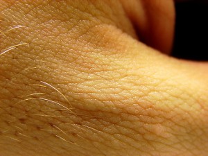
Really. Come on. Who isn’t interested in hair? Hair growth, hair loss, hair thickness, hair shape, hair location. I’d bet that everyone of us spends at least a minute or two each day thinking about (or, if you’re like me, futilely plucking and prodding at) the state of our locks.

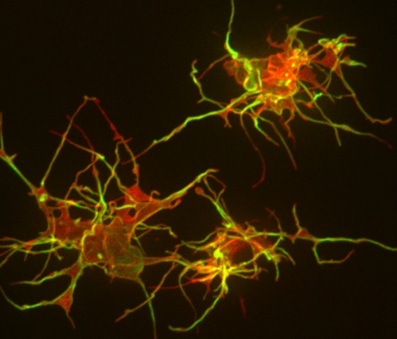
Now Stanford researchers have delved deep into the cells surrounding our hair follicles to better understand what makes them grow and maintain hair. Perhaps not surprisingly, the answer lies in the stem cells (here, called ‘bulge cells’) within the follicle.

Specifically, research associate Yiqin Xiong, PhD, and associate professor of medicine Ching-Pin Chang, MD, PhD, have identified a signaling circuit that controls the cells’ activity. The research was published yesterday in Developmental Cell (subscription required). As Chang explained in an e-mail to me:

By promoting self-renewal of stem cells, this circuit maintains a healthy pool of bulge cells for repeated cycles of hair growth and regeneration. Each cycle of hair regeneration is initiated by the activation of this circuit in those bulge cells, and subsequent growth of the hair is sustained by the circuit in hair matrix cells. Besides hair regeneration, the circuit is triggered by skin injury to stimulate migration of the bulge cells to the wounded area to differentiate into epidermal cells, thereby regenerating epidermis over the wounded skin.

In the past, news about hair growth (and how to stimulate it) has been a trigger for a deluge of interest from media and individuals struggling with… (how shall we say it?) ‘hair problems.’ But the research has many implications beyond hair, or the lack thereof. For example, the presence or absence of hair follicles on the skin affect how the skin heals after a wound, and whether a scar remains. According to Chang:

This molecular circuit in the hair follicle can be targeted for therapeutic purposes. Because of its activity in hair regeneration, inhibition of this circuit can reduce hair growth in patients with excessive hairiness (hirsutism), whereas activation of this pathway can promote hair growth for people with baldness (alopecia). Also, for its activity during epidermal regeneration, activation of the circuit can facilitate wound healing for patients receiving surgery and for diabetic patients who have wounds that are difficult to treat. The activity of the circuit in both hair follicle and epidermal regeneration may have additional therapeutic benefit. Lack of hair follicles in a wounded area is a hallmark of scar formation. Targeting this pathway has the advantage of promoting both hair follicle formation and wound repair, thus reducing scar formation in the wound.

Interestingly, one of the key molecules, called Brg1, involved in this regulatory circuit has also been implicated in previous work from Chang’s lab in the enlargement of the heart and in fetal heart development. It’s apparent this story has many layers, some more than skin deep.

Previously: Examining the role of genetics in hair loss and Epigenetics: the hoops genes jump through,
Photo by Furryscaly

Imaging, In the News, Neuroscience, Research, Stanford News

Neuroscience studies often underpowered, say researchers at Stanford, Bristol

neuroscience-studies-often-underpowered-say-researchers-at-stanford-bristol

I’m ashamed to admit that the study of statistics was regarded (at least by me) as a necessary evil when I was in graduate school. I vaguely remember one course that attempted to teach a lecture hall of sleepy, stressed-out students how to calculate p values, the differences between retrospective, prospective and case-control studies, and the nuances between sensitivity and specificity. And don’t even get me started on odds ratios. Can you tell I’m still a bit fuzzy? In fact, I keep a reference guide at my desk for help (which I have to consult embarrassingly often).

Statistics might be dull, but there’s no denying its importance in scientific research - and the fallout when scientists fail to appreciate its power. Now, Stanford researcher John Ioannidis, MD, DSci, (of the “Why most published research findings are false” fame) has joined forces with Marcus Munafo, PhD, and others at the University of Bristol to publish a new study in in Nature Reviews Neuroscience (subscription required) delineating the statistical flaws in many published neuroscience studies. Essentially, the researchers found that, although many scientists realize that an under-powered study (for example, one with too few study subjects to adequately capture the phenomena being investigated) is less likely to find statistically significant results, they don’t necessary realize the converse: that any statistically significant finding from such a study is less likely to represent a true effect.

Stellar science blogger Ed Yong explains the sobering implications in an excellent post today:

Statistical power refers to the odds that a study will find an effect—say, whether antipsychotic drugs affect schizophrenia symptoms, or whether impulsivity is linked to addiction—assuming those effects exist. Most scientists regard a power of 80 percent as adequate—that gives you a 4 in 5 chance of finding an effect if there’s one to be found. But the studies that Munafo’s team examined tended to be so small that they had an average (median) power of just 21 percent. At that level, if you ran the same experiment five times, you’d only find an effect on one of those. The other four tries would be wasted.

But if studies are generally underpowered, there are more worrying connotations beyond missed opportunities. It means that when scientists do claim to have found effects—that is, if experiments seem to “work”—the results are less likely to be real. And it means that if the results are actually real, they’re probably bigger than they should be. As the team writes, this so-called “winner’s curse” means that “a ‘lucky’ scientist who makes the discovery in a small study is cursed by finding an inflated effect.”

I encourage you to read all of Ed’s post, which includes multiple comments from Ioannidis, Munafo and other researchers uninvolved in the study. It’s a fascinating analysis of why many studies are designed as they are, and it discusses some of the obstacles that must be overcome to improve their fidelity. And don’t overlook the comment stream, which is currently hosting a rich discussion among scientists in the field.

Previously: NIH funding mechanism “totally broken” says Stanford researcher, Research shows small studies may overestimate the effects of many medical interventions and Animal studies: necessary but often flawed, says Stanford’s Ioannidis
Photo by futureshape

CDC, Global Health, In the News, Public Health

H7N9 got you aflutter? Wired offers help sorting fact from fiction

I admit to a certain sense of mounting dread about the news of the new H7N9 influenza virus arising in China. And the never-ending supply of Tweets (alarmist and otherwise) are not helping one little bit. That’s why I appreciated this article posted today by Wired reporter and author Maryn McKenna (she’s sometimes referred to as Scary Disease Girl, due to her focus on global health and infectious diseases).

McKenna breaks the current news down into a quick primer, based on her past experiences reporting on that ‘other bird flu’ H5 N1 (remember that one?) ten years ago. She follows with a caution to beware- or at least to be aware- of the sources of news of this quickly moving story, and an explanation of some peculiarities in Chinese media that may hamper or distort reporting. She also draws a parallel between what’s happening now with H7N9 and H5N1- pointing out that the latter never erupted in humans as it was first feared. Says McKenna:

And H7N9 might not, as well. It is far too soon to say, despite the rapidly escalating case count and the reports — which came in while I was writing this — of a possible animal reservoir in pigeons and a possible human-to-human case. I have been writing about flu and possible pandemics since 1997 — for what it’s worth, I wrote the first story in the US in 1997 about that first H5N1 case in Hong Kong — and so at this early point, what I most want to say is this: We all love scary diseases. (If you didn’t, you wouldn’t be reading this blog.) But there is a fog of war in disease emergencies, just as there is in military ones, and it is very easy to get lost in it.

It will take a while for this story to become more clear. Anticipating that, I want to suggest some things to think about as you follow the news.

She ends with this great advice:

[...] Don’t assume that everyone who is loading information onto their blogs or pushing it onto Twitter is doing it in a sharing spirit of helpfulness. There are people — you can see this already — who are opportunistically using this to feed their egos, angle for jobs, or generally to stir up trouble. More than ever, it’s important to be skeptical about the sources of the information you consume.

McKenna makes it easier for us to practice what she preaches by listing several reputable news sources-traditional, web-based and, even on, Twitter- that should be reliable sources of information. You can follow McKenna on Twitter at @marynmck.

Previously: “Superbug” author discusses dangers, history and treatment of MRSA and Image of the week: What H5N1 looks like

Neuroscience, Research, Stanford News

Amyloid, schmamyloid: Stanford MS expert finds dreaded proteins may not be all bad

amyloid-schmamyloid-stanford-ms-expert-finds-dreaded-proteins-may-not-be-all-bad

I have to thank my colleague Bruce Goldman for taking some time off recently and leaving me to tackle a story that’s normally in his territory of neurobiology. The research article he handed off is a fascinating extension of a biological plotline (subscription required) that Bruce first wrote about last August in Science Translational Medicine. The idea is that, although a particular protein called beta amyloid has for years been considered to be the smoking gun in Alzheimer’s disease, a Stanford neurologist found that it’s actually beneficial in animal models of multiple sclerosis.

Now, Science Translational Medicine has published the latest iteration of the researcher’s work, showing that small pieces of several other amyloid-forming proteins can also relieve symptoms in mice with MS. From our release:

“What we’re finding is that, at least under certain circumstances, these amyloid peptides actually help the brain,” said Lawrence Steinman, MD, professor of neurology and neurological sciences and of pediatrics. “This really turns the ‘amyloid-is-bad’ dogma upside down. It will require a shift in people’s fundamental beliefs about neurodegeneration and diseases like multiple sclerosis, Alzheimer’s and Parkinson’s.”

[...] Taken together, the studies begin to suggest the radical new idea that full-length, amyloid-forming proteins may in fact be produced by the body as a protective, rather than destructive, force. In particular, Steinman’s study shows that these proteins may function as molecular chaperones, escorting and removing from sites of injury specific molecules involved in inflammation and inappropriate immune responses.

This remarkable rehabilitation of the black sheep of neurobiology was great fun to write, and could change how neurobiologists think about and attempt to treat neurodegeneration. For example, it’s possible that therapies aimed at removing all amyloid-forming proteins from the body may hinder, rather than help, attempts to treat a patient’s symptoms.

Steinman is currently scheduled (barring other breaking news) to speak about his findings on April 5 (around noon Pacific time) with Ira Flatow, the host of NPR’s popular Science Friday program. I’m sure Bruce and I will both be tuning in to hear more about this fascinating story. We may have to duke it out, though, when it comes to writing the next press release about Steinman’s research…

Previously: Black hat in Alzheimer’s, white hat in multiple sclerosis?, Brain sponge: Stroke treatment may extend time to treat brain damage and Stanford neuroimmunologist discusses recent advances in MS research
Photo of Lawrence Steinman by Steve Fisch

Research, Stanford News

Mast cells not required for wound healing, according to Stanford study

mast-cells-not-required-for-wound-healing-according-to-stanford-study

Next time you get a paper cut, pause for a moment to consider the molecular cast of characters racing to the wound site. Proper healing requires an intricate dance between many cell types, and - for researchers and clinicians interested in tissue repair - ruling out the red herrings in the process is just as important as identifying the most-critical players.

Yesterday, Stanford stem-cell scientist and pediatric plastic and reconstructive surgeon Michael Longaker, MD, and pathologist Stephen Galli, MD, published a study in PLoS ONE showing that one class of cells, called mast cells, previously thought to be important in healing is actually not required. As Longaker explained in an e-mail to me:

Wound repair is a complex biologic process involving the interaction of many cell types to replace the lost or damaged tissue. The order in which cells arrive at the wound has been extensively studied and is generally thought to be neutrophils followed by macrophages, followed by fibroblasts, and finally mast cells. The role of each of these cells has been studied with the exception of mast cells, and very little is known about how they function in wound repair. Our study examines the role of mast cells using three mast-cell deficient mouse models. Our results indicate that mast cells do not play a significant non-redundant role during excisional wound repair.

The upshot? It appears that mast cells, which are heavily involved in allergic responses, function more like ambulance chasers than emergency responders when skin meets knife. Or, as in the case above, paper.

Previously: A good ‘coach’ and the right environment keeps stem cells in check, say Stanford researchers and A scar-free future for kids?

Stanford Medicine Resources: