Published by
Stanford Medicine

Category

Neuroscience

Behavioral Science, LGBT, Neuroscience, Sexual Health, Stanford News

Distinction with a difference: Transgendered neurobiologist picked for National Academy of Science membership

distinction-with-a-difference-transgendered-neurobiologist-picked-for-national-academy-of-science-membership

The National Academy of Sciences recently celebrated its 150th birthday by, among other things, conferring membership on Ben Barres, MD, PhD. Additional NAS admittees from Stanford were sleep scientist Emmanuel Mignot, MD, PhD, and bioengineer Steve Quake, PhD.

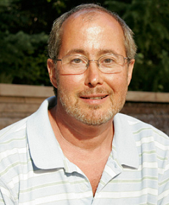
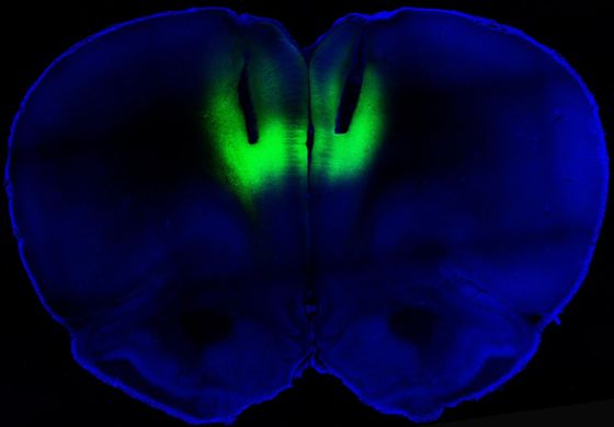
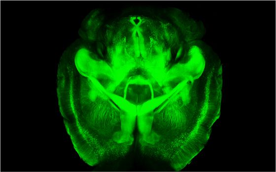
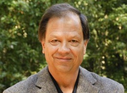
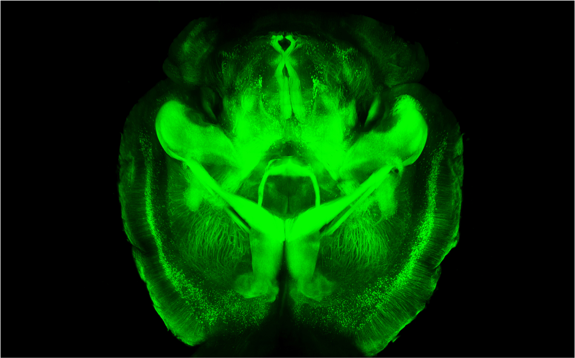
A distinguished scientist by anybody’s yardstick, as well as the chair of Stanford’s ironically named neurobiology department, Barres is a leading light in the study of glial cells (collectively known as glia), the 90 percent of all the cells in the brain that aren’t nerve cells.

The term”glia” is derived from the Greek word for glue. Like Rodney Dangerfield, glial cells once got no respect. They were thought of, in fact, as not much more than “brain glue”: mere structural scaffolds for the organ’s much more revered nerve cells.

Barres’ research has proved that hypothesis incorrect, to say the least. (For details, click here.) Discoveries coming out of his lab include, to name one example, glial cells’ crucial role in determining exactly when and where nerve-cell connections in the brain are made, tweaked to strengthen or weaken them, or destroyed.

You don’t get much more respectable than that: Those connections pretty much define the thoughts we have, the emotions and sensations we experience and the actions we take.

The man who, as much as anyone, has brought a set of unsung cells a newly elevated status would like to see another group get more respect: the estimated 0.3 percent of Americans who are transgender.

“I’m the first transgender scientist to make into the National Academy of Science,” says Barres, who began life under another first name: Barbara.

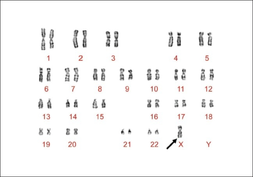
“We don’t know if other members past or present are or were transgender,” demurs an NAS representative. And after all, how would they? What kind of statistics could be compiled by an organization that doesn’t ask or track the sexual orientations, much less the gender identities, of its membership? Who would have even considered asking such a question 20 or 30 years ago, much less running sex-chromosome tests on cheek swabs from prospective, current or posthumous members?

But it’s a pretty safe bet that if any previously admitted NAS member were openly transgender, we’d have heard about it. (Transgendered computer scientist Lynn Conway was admitted to the National Academy of Engineering in 1989.)

One is tempted to compare Barres to Jackie Robinson, who broke the Major League Baseball’s color barrier in 1947 - except that the latter had to put up with a whole lot more grief from his fellow major-league ballplayers than Barres is likely to encounter from his peers.

“We heartily congratulate Prof. Barres on his election,” says NAS spokesperson Bill Skane.

In science, if anywhere, diverse perspectives drive innovation. ”Don’t ever let anyone make you feel bad about being different,” Barres tells young scientists. “Your difference is your greatest advantage.”

Previously: Malfunctioning glia - brains cells that aren’t nerve cells - may contribute big time to ALS and other neurological disorders, Neuroinflammation, microglia, and brain health in the balance and Unsung brain-cell population implicated in variety of autism

Ask Stanford Med, Autism, Neuroscience, Parenting, Pediatrics, Research, Stanford News

Ask Stanford Med: Director of Stanford Autism Center taking questions on research and treatment

ask-stanford-med-director-of-stanford-autism-center-taking-questions-on-research-and-treatment

Among school-aged children in the United States an estimated one in 50 has been diagnosed with autism spectrum disorder, according to a recent survey (.pdf) from the Centers for Disease Control and Prevention. In addition to raising concerns among researchers and parents about why the number of cases has increased, the findings underscored the need to do more autism research and to provide support and services for families caring for autistic children.

To help parents and others in the local community better understand the growing prevalence of autism and to learn about treatments and research advancements, the Stanford Autism Center at Packard Children’s Hospital will host its sixth annual Autism Spectrum Disorders Update on June 1. The event offers an opportunity for exchange between parents, caregivers and physicians and provides an overview of the center’s clinical services and ongoing autism research at the School of Medicine.

In anticipation of the day-long symposium, we’ve asked Carl Feinstein, MD, director of the center, to respond to your questions about issues related to autism spectrum disorder and to highlight how research is transforming therapies for the condition.

At the Stanford Autism Center, Feinstein works with a multidisciplinary team to develop treatments and strategies for autism spectrum disorders. In providing care and support for individuals with autism and their families, Feinstein and colleagues identify ways of targeting the primary autism symptoms, while also paying attention to associated behavior problems that may hold a child back from school or community involvement or seriously disrupt family life.

Questions can be submitted to Feinstein by either sending a tweet that includes the hashtag #AskSUMed or posting your question in the comments section below. We’ll collect questions until Wednesday (May 15) at 5 PM Pacific Time.

When submitting questions, please abide by the following ground rules:

  • Stay on topic
  • Be respectful to the person answering your questions
  • Be respectful to one another in submitting questions
  • Do not monopolize the conversation or post the same question repeatedly
  • Kindly ignore disrespectful or off topic comments
  • Know that Twitter handles and/or names may be used in the responses

Feinstein will respond to a selection of the questions submitted, but not all of them, in a future entry on Scope.

Finally – and you may have already guessed this – an answer to any question submitted as part of this feature is meant to offer medical information, not medical advice. These answers are not a basis for any action or inaction, and they’re also not meant to replace the evaluation and determination of your doctor, who will address your specific medical needs and can make a diagnosis and give you the appropriate care.

Previously: New public brain-scan database opens autism research frontiers, New autism treatment shows promising results in pilot study, Autism’s effect on family income, Study shows gene mutation in brain cell channel may cause autism-like syndrome, New imaging analysis reveals distinct features of the autistic brain and Research on autism is moving in the right direction
Photo by Wellcome Images

Genetics, Neuroscience, Pediatrics, Research, Stanford News

Does it matter which parent your “brain genes” came from?

does-it-matter-which-parent-your-brain-genes-came-from

Does it make a difference if a gene – or group of genes – is inherited from your mother or your father?

That’s the question behind the study of genomic imprinting, a phenomenon in which a small percent of genes are thought to be expressed differently depending on which parent they came from. In particular, animal research suggests imprinting may affect aspects of brain development. Researchers wonder if genomic imprinting might explain differences in brain anatomy seen between men and women, such as men’s larger brain volumes.

A new Stanford study, published today in the Journal of Neuroscience, adds to evidence that genomic imprinting is, in fact, happening in humans’ brains. The finding comes from MRI brain scans performed on a group of young girls with Turner syndrome, a chromosomal disorder in which a girl or woman has one missing or malfunctioning X chromosome. Turner syndrome gives an unusual opportunity to study genetic imprinting, because it allows comparisons of individuals who received a single X from Mom to those who got a single X from Dad. (The typical two-X-chromosome female body expresses a mosaic of Mom’s X and Dad’s X, making it impossible to tease apart the effects of the two parents. Males invariably get their single X chromosome from their mothers, so their cells always express the maternal X.)

The Stanford team, led by Allan Reiss, MD, documented several distinctions between the brains of Turner syndrome girls who have only a maternal X, those with only a paternal X, and typical girls with two X chromosomes, such as differences in the thickness and volume of the cortex, and in the surface area of the brain. The work helps clarify murky results from earlier studies of adults with Turner syndrome, the researchers say, because many adult women with Turner syndrome take estrogen supplements, which may have their own effects on brain development. None of the girls in the new study had taken estrogen.

The most tantalizing part of the paper is the scientists’ comment on the implications of their work for our general understanding of genetic imprinting. In part, they say:

By far, the most consistent finding with regard to sex differences in brain anatomy is the larger brain volume found in males compared with females. Although our groups did not differ on most whole-brain measures, our analyses revealed the existence of significant trends on total brain volume, gray matter volume and surface area, where these variables increased linearly from the Xp [paternal X] group being smallest, to the Xm [maternal X] group being largest, with typically developing girls in between. Considering that typically developing males invariably inherit the maternal X chromosome, while typically developing females inherit both and randomly express one of them in each cell, a linear increase in brain volume as seen in the present study is in agreement with what would be expected if imprinted genes located on the X chromosome were involved in brain size determination.

In other words, men may have their mothers to thank for their larger brains.

Karyotype image from a Turner Syndrome patient by S Suttur M, R Mysore S, Krishnamurthy B, B Nallur R - Indian J Hum Genet (2009).

Neuroscience, Podcasts, Stanford News

Brain’s gain: Stanford neuroscientist discusses two major new initiatives

brains-gain-stanford-neuroscientist-discusses-two-major-new-initiatives

The brain has gotten a lot of attention lately. Last month, President Obama announced a $100 million decades-long research initiative to “unlock”, as he called it, “the mystery of the three pounds of matter that sits between the ears.” And, in the arena of jaw-dropping science, Stanford’s Karl Diesseroth, MD, PhD, and Kwanghun Chung, PhD, recently unveiled CLARITY - a process that rendered a mouse brain transparent. Thomas Insel, MD, director of the National Institute of Mental Health, called the Stanford researchers’ work, “frankly spectacular.”

Primed for this moment of brain fame is Stanford’s Bill Newsome, PhD, who has been toiling in the field of neuroscience for nearly three decades. His international renown as a research scientist catapulted him to two new key brain posts: vice chair of the federal BRAIN initiative and director of a new interdisciplinary neuroscience institute at Stanford.

I talked with Newsome about both efforts for my latest 1:2:1 podcast. I began by asking him, “Why now?” What has propelled the brain to the front of the food chain in federal funding?

He called the Obama administration “prescient” for putting forth the federal effort. “There has never been a bigger moment of progress for brain research than there is now,” he told me. He describes this time as a “tipping point” where putting the pedal on the accelerator will make a whole new world of research possible.

Newsome is also cautious. Over-promising breakthroughs is clearly not in his vernacular. Yet he see this moment with clarity and admits that accelerating what we’re already doing will allow us “to get new data about the brain that we never dreamed possible.”

Previously: Co-leader of Obama’s BRAIN Initiative to direct Stanford’s interdisciplinary neuroscience institute, Lightning strikes twice: Optogenetics pioneer Karl Deisseroth’s newest technique renders tissues transparent, yet structurally intact, Experts weigh in on the new BRAIN Initiative and A federal push to further brain research

Aging, Chronic Disease, Neuroscience, Research, Stanford News

Revealed: The likely role of Parkinson’s protein in the healthy brain

revealed-the-likely-role-of-parkinsons-protein-in-the-healthy-brain

So-called Lewy bodies - gumball-like clumps rich in a mystery molecule called alpha-synuclein - abound in Parkinson patients’ brains and are considered the hallmark of the disease. Up to now, researchers have had few solid clues as to what this “black hat” protein is doing in the brain in the first place.

But a team led by Stanford neuroscientists Tom Sudhof, MD, and Axel Brunger, PhD, has revealed a likely critical role played by alpha-synuclein in healthy brains. Their discovery is described in an article just published in the open-access online journal eLife.

Each of the human brain’s roughly 200 billion nerve cells communicates directly with, on average, 10,000 others by squirting signaling chemicals called neurotransmitters at them. It is all this squirting that underpins our thoughts, feelings and movements.

Of course, the brain’s activity is no mob squirt-gun shootout. Consider: The 2 quadrillion separate nerve-cell connections in your brain or mine roughly equal the number of stars in 7,000 Milky Way galaxies. For our most exalted organ to do its job, the signals that nerve cells send must be marked by profound precision, both in their intensity and in their timing.

As I wrote in my release accompanying the eLife article:

Nerve cells don’t simply squirt out neurotransmitters willy-nilly. Within the complex networks that constitute our brains, every individual nerve cell has a lengthy, snaking, tubular extension cord, or axon, that hooks up with thousands of other nerve cells. Neurotransmitters are housed within tiny bubble-like packets in the cell. These packets congregate in myriad small, bulbous nozzles dotting the axon, with each bulb abutting a downstream nerve cell. When an electrical impulse travels down the axon on which those bulbs reside, it triggers the fusion of the neurotransmitter-packed packets with the nerve cell’s outer membrane. The packets’ contents then spill into the narrow space separating the bulbs from the nerve cells they abut.

The Sudhof-and-Brunger team was able to show that alpha-synuclein helps regulate the orderly clustering of the neurotransmitter-loaded packets near their release sites. Alpha-synuclein has to be present in the right amounts, though; too much or too little has untoward consequences - which could explain why previous research has yielded conflicting results.

It’s nice to know, before messing around with it in living people, that in the healthy brain alpha-synuclein is a lot more than just a raw material in a gumball factory. Drug companies may have perhaps been led down some blind alleys as a result of locking in, too early, on the notion that yet another clump-generating protein, A-beta, was the Bad Guy in Alzheimer’s disease and that, it followed, getting rid of it would be a good idea. Maybe not so fast

Previously: Nervous breakdown: Preventing demolition of faulty proteins counters neurodegeneration in lab mice, Stanford scientist sets sail on new publishing model with launch of open-access, embargo-free journal and Stanford study identifies molecular mechanism that triggers Parkinson’s
Photo by akeg

Imaging, Neuroscience, Pediatrics, Research, Science, Stanford News

Peering into the brain to predict kids’ responses to math tutoring

peering-into-the-brain-to-predict-kids-responses-to-math-tutoring

Third grade is a critical year for learning arithmetic facts, but while math comes easily to some children, others struggle to master the basics.

Now, researchers at Stanford have new insight into what separates adept young math students from those who have difficulty. The difference, described in a paper published today in the Proceedings of the National Academy of Sciences, can’t be detected with traditional intelligence measures such as IQ tests. But it shows up clearly on brain scans, as the new study’s senior author explained in our press release:

“What was really surprising was that intrinsic brain measures can predict change — we can actually predict how much a child is going to learn during eight weeks of math tutoring based on measures of brain structure and connectivity,” said Vinod Menon, PhD, the study’s senior author and a professor of psychiatry and behavioral sciences.

Menon’s research team conducted structural and functional MRI brain scans before third-grade students received 8 weeks of individualized math tutoring. The tutoring followed a well-validated format, combining instruction on math concepts with practice of math problems emphasizing speed. All the children who received math tutoring improved their math performance, but the performance improvements varied a lot — from 8 percent to 198 percent.

A few specific brain characteristics were particularly good at predicting which kids would benefit most from tutoring. In particular, a larger and better-wired hippocampus predicted performance improvements. The brain structures highlighted in the study are implicated in forming memories, and differ from the portions of the brain that adults use when they are learning about math. The fact that these systems are involved helps to explain why the combination of conceptual explanations and sped-up practice that the study’s tutors used is effective, Menon explained:

“Memory resources provided by the hippocampal system create a scaffold for learning math in the developing brain,” Menon said. “Our findings suggest that, while conceptual knowledge about numbers is necessary for math learning, repeated, speeded practice and testing of simple number combinations is also needed to encode facts and encourage children’s reliance on retrieval — the most efficient strategy for answering simple arithmetic problems.” Once kids are able to pull up answers to basic arithmetic problems automatically from memory, their brains can tackle more complex problems.

Next, the researchers plan to examine how brain wiring changes over the course of tutoring. The new findings could also help educators understand the basis for math learning disabilities, and may even provide a foundation for figuring out what kind of instruction could help children overcome these problems.

Previously: New research tracks “math anxiety” in the brain and We’ve got your number: Exact spot in brain where numeral recognition takes place revealed
Photo by Canadian Pacific

Image of the Week, Neuroscience

Image of the Week: Prelimbic cortex illuminated with optogenetic stimulation

image-of-the-week-prelimbic-cortex-illuminated-with-optogenetic-stimulation

Researchers at the National Institutes of Health and University of California-San Francisco have found that stimulating a key part of the brain reduces compulsive cocaine-seeking and suggests the possibility of changing addictive behavior generally. NIH Director Francis Collins, MD, PhD, discussed the study, and the significance of the findings in a blog post earlier this month:

The researchers studied rats that were chronically addicted to cocaine. Their need for the drug was so strong that they would ignore electric shocks in order to get a hit. But when those same rats received the laser light pulses, the light activated the [prelimbic area of the prefrontal cortex], causing electrical activity in that brain region to surge. Remarkably, the rat’s fear of the foot shock reappeared, and assisted in deterring cocaine seeking. On the other hand, when the team used a different optogenetics technique to reduce activity in this same brain region, rats that were previously deterred by the foot shocks became chronic cocaine junkies.

Clearly this same approach wouldn’t be used in humans. But it does suggest that boosting activity in the prefrontal cortex using methods like transcranial magnetic stimulation (TMS), which is already used to treat depression, might help.

This image shows optogenetic stimulation using laser pulses illuminating the prelimbic cortex. The channelrhodopsins used to create the photo were provided to researchers by Stanford bioengineer Karl Deisseroth, MD, PhD.

Previously: Better than the real thing: How drugs hot wire our brains’ reward circuitry, The brain’s control tower for pleasure and Addiction: All in the mind?
Photo by Billy Chen and Antonello Bonci

In the News, Mental Health, Neuroscience

How the brain processes trauma and why support, altruism can ease fear

how-the-brain-processes-trauma-and-why-support-altruism-can-ease-fear

The traumatic events at yesterday’s Boston Marathon have many of us bracing ourselves for what might be coming next. And, as explained in a Healthland piece, this feeling of being on high alert is a result of how our brain processes traumatic experiences.

As writer Maia Szalavitz explains, “when the brain is under severe threat, it immediately changes the way it processes information, and starts to prioritize rapid responses.” While this behavior is important to our survival, it can be be harmful to our health if it persists after the threat has passed. So what can we do to help each other heal from the tragedy and reduce the risk of those most affected from developing post-traumatic stress disorder (PTSD)? Szalavitz writes:

Fortunately, our brains are designed to modulate fear responses and at least 80% of people exposed to a severe traumatic event will not develop PTSD. Studies show that the more support, altruism and connection people share, the lower the risk for the disorder and the easier the recovery. Because such interactions aren’t always easy in the immediate aftermath of a harrowing experience, Hollander is investigating whether medications based on oxytocin— a hormone linked with love and parent/child bonding— might help to ease this connection.

If fear short circuits the brain’s normally logical and reasoned thinking, social support may be important in rerouting those networks back to their normal state. Which is why the selflessness and altruism we see in the wake of terror attacks is often the key to helping us to process and overcome the shock of living through them.

Szalavitz’s message of using compassion to combat fear was echoed in this TED blog post, which encourages people to “look for the helpers” as we process what happened yesterday, and in Mashable’s list of touching acts of kindness at the marathon.

Previously: Can social media improve the mental health of disaster survivors?, Grieving on Facebook: A personal story and 9/11: Grieving in the age of social media
Photo by Alex E. Proimos

Behavioral Science, Evolution, Neuroscience, Research, Stanford News

We’ve got your number: Exact spot in brain where numeral recognition takes place revealed

weve-got-your-number-exact-spot-in-brain-where-numeral-recognition-takes-place-revealed

Your brain and my brain are shaped slightly differently. But, it’s a good bet, in almost the identical spot within each of them sits a clump of perhaps 1 to 2 million nerve cells that gets much more excited at the sight of numerals (“5,” for example) than when we see their spelled-out equivalents (“five”), lookalike letters (“5″ versus “S”) or scrambled symbols composed of rearranged components of the numerals themselves.

Josef Parvizi, MD, PhD, director of Stanford’s Human Intracranial Cognitive Electrophysiology Program, and his colleagues identified this numeral-recognition module by recording electrical activity directly from the brain surfaces of epileptic volunteers. Their study describing these experiments was just published in The Journal of Neuroscience.

As I explained in my release about the work:

[A]s a first step toward possible surgery to relieve unremitting seizures that weren’t responding to therapeutic drugs, [the patients had] had a small section of their skulls removed and electrodes applied directly to the brain’s surface. The procedure, which doesn’t destroy any brain tissue or disrupt the brain’s function, had been undertaken so that the patients could be monitored for several days to help attending neurologists find the exact location of their seizures’ origination points. While these patients are bedridden in the hospital for as much as a week of such monitoring, they are fully conscious, in no pain and, frankly, a bit bored.

Seven patients, in whom electrodes happened to be positioned near the area Parvizi’s team wanted to explore, gave the researchers permission to perform about an hour’s worth of tests. In the first, they watched a laptop screen on which appeared a rapid-fire random series of letters or numerals, scrambled versions of them, or foreign number symbols with which the experimental subjects were unfamiliar. In a second test, the experimental subjects viewed, again in thoroughly mixed-up sequence, numerals along with words for them as well as words that sounded the same (1″, “one”, “won”, “2″, “two”, “too”, etc.).

A region within a part of the brain called the inferior temporal gyrus showed activity in response to all kinds of squiggly lines, angles and curves. But within that area a small spot measuring about one-fifth of an inch across lit up preferentially in response to numerals compared with all the other stimuli.

The fact that this spot is embedded in a larger brain area generally responsive to lines, angles, and curves testifies to the human brain’s “plasticity:” its ability to tailor its form and function according to the dictates of experience.

“Humans aren’t born with the ability to recognize numbers,” says Parvizi. He thinks evolution may have generated, in the brains of our tree-dwelling primate ancestors, a brain region particularly adept at computing lines, angles and curves, facilitating snap decisions required for swinging quickly from one branch to the next.

Apparently, one particular spot within that larger tree-branch-interesection recognition area is easily diverted to the numeral-recognition activity constantly rewarded by parents and teachers during the numeracy boot camp called childhood.

Nobody can say those little monkeys don’t learn anything in kindergarten.

Previously: Metamorphosis: At the push of a button, a familiar face becomes a strange one and Why memory and math don’t mix: They require opposing states of the same brain circuitry
Photo by qthomasbower

Image of the Week, Imaging, Neuroscience, Research, Stanford News

Image of the Week: 3-D rendering of a clarified brain

image-of-the-week-3-d-rendering-of-a-clarified-brain

Earlier this week, fellow Scope contributor Bruce Goldman reported on a paradigm-shifting process developed by Stanford psychiatrist and bioengineer Karl Deisseroth, MD, PhD, and colleagues. Using the process, called CLARITY, scientists were able to turn a mouse brain into an “optically transparent, histochemically permeable replica of itself.”

National Institutes of Health Director Francis Collins, MD, PhD, commented on the breakthrough in a recent blog post, saying:

CLARITY is powerful. It will enable researchers to study neurological diseases and disorders, focusing on diseased or damaged structures without losing a global perspective. That’s something we’ve never before been able to do in three dimensions.

This haunting image depicts a three-dimensional rendering of clarified brain imaged from the ventral half. To fully experience the new method’s awe-inspiring capabilities, watch this fly-through video.

Previously: Scientific community (and Twitter) buzzing over Stanford’s see-through brain, Lightning strikes twice: Optogenetics pioneer Karl Deisseroth’s newest technique renders tissues transparent, yet structurally intact and Peering deeply – and quite literally – into
Photo by Deisseroth lab

Stanford Medicine Resources: