 Stem-cell therapy for damaged hearts is a brilliant idea whose time has not yet come. To date, human and animal trials - there have been quite a few - in which stem cells were injected into cardiac tissue to treat severe heart attacks or substantial heart failure have mostly produced poor results.
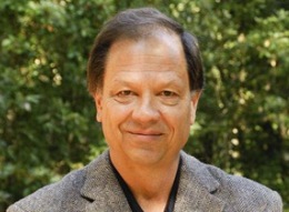
Stanford’s Sam Gambhir, PhD, MD, who heads the medical school’s Department of Radiology, thinks he knows part of the reason why, and he may have found a way around it.
At present, there’s no way to ensure against faulty initial placement, he told me in an interview about his study describing the proposed solution, just published in Science Translational Medicine. “You can use ultrasound to visualize the needle through which you deliver stem cells to the heart. But once those cells leave the needle, you’ve lost track of them.”
In my release about the work, I wrote:
As a result, key questions go unanswered: Did the cells actually get to the heart wall? If they did, did they stay there, or did they diffuse away from the heart? If they got there and remained there, for how long did they stay alive? Did they replicate and develop into heart tissue?
Gammbhir’s lab has figured out a way to “mark” stem cells before infusing them into the heart, rendering them visible to standard ultrasound as they’re squeezed out of the needle. The key was to invent an innovative imaging agent, in the form of nanoparticles whose diameters clustered just below one-third of a micron — less than one-three-thousandth the width of a human hair. The nanoparticles’ main ingredient, silica, shows up on ultrasound. The particles were also doped with the rare-earth element gadolinium, so they can also be observed using MRI.
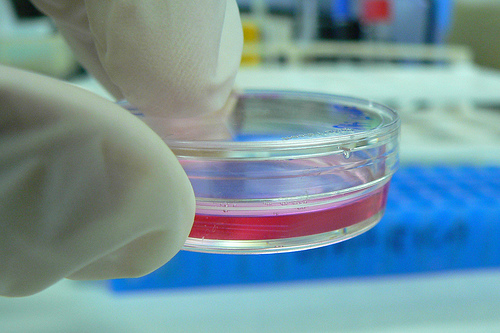
It turns out that mesenchymal stem cells — a class of cells often used in heart-regeneration research because they can differentiate into beating heart cells and because they can sometimes be harvested directly from the patients themselves, avoiding possible immune-compatibility problems — were happy to gobble up the nanoparticles in a lab dish without losing any of their ability to survive, thrive, and replicate themselves.
When Gambhir and his associates infused these nanoparticle-loaded stem cells into the hearts of healthy mice, they were indeed able to monitor the cells via ultrasound after they left the needle tip, guide them to the targeted area of the heart wall, and still get a strong MRI signal from the cells two weeks later.
Stem-cell therapy for damaged hearts isn’t going to be cheap anytime soon. (A wild guess of, say, $50,000 per procedure is probably not too far off the mark.) But a one-time delivery, if it worked, could replace a lifetime of constant drug administration. Adding what Gambhir estimates might be another $2,500 a pop for the added imaging capability is likely to be hugely cost-effective, because it could greatly improve the odds of the procedure’s success.
Previously: Nanoparticles home in on human tumors growing in mice’s brains, increase accuracy of surgical removal, Nanomedicine moves ones step closer to reality and Developing a new molecular imaging system and technique for early disease detection
Photo by miguelb