Published by
Stanford Medicine

Author

Medical Education, Science, Stanford News

Stanford’s RISE program gives high-schoolers a scientific boost

stanfords-rise-program-gives-high-schoolers-a-scientific-boost

While a solution to low test scores and lackluster interest in science and engineering careers among schoolchildren nation-wide remains elusive, outreach initiatives can effect change at the local level. A Stanford Report article today describes the university’s Raising Interest in Science and Engineering (RISE) program, which matches select low-income and under-represented minority high-school students with expert guidance, work experience in laboratories, and exposure to a variety of academic and industry experiences. From the piece:

In many cases, minority and lower-income students who are particularly unprepared for and underrepresented in the sciences don’t even see a science or engineering degree as an option.

….

“These are kids who may not have scientists or engineers in their networks,” said Kaye Storm, MA, director of Stanford’s Office of Science Outreach, which runs RISE. “They know they like science or engineering, but they don’t really know what that might mean in terms of an internship or career.”

By exposing the students to an academic laboratory environment and introducing them to potential scientific contacts, RISE aims to bridge that gap between student talent and access to a college degree in the sciences.

All 89 students who have participated in RISE since the program’s 2006 inception have gone on to college or will enter it this year, and 12 have attended Stanford. RISE alumna Alison Logia tells writer Max McClure:

“In school, I’d end up in science classes with predesigned labs,” said Logia. “But when I came to Stanford it was different. When you get your results, you can’t look them up in a book to see if they’re correct, because no one’s ever done this experiment before.”

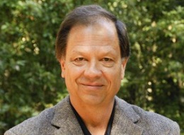
Logia, a graduate of Sequoia High School in Redwood City, worked for two summers in the lab of chemical engineering Professor Gerald Fuller [PhD]. Although she knew she was interested in math and science in high school, her experiences in the Fuller Lab taught her “how to work in a lab, how to talk to a professor,” and solidified her desire to go into engineering.

Previously: Stanford science program for teens receives Presidential Award, I know what you did this summer: High-school interns share their experiences at Stanford, A look at the Stanford Medical Youth Science Program and A prescription for improving science education
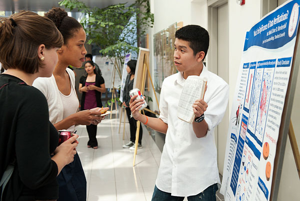
Photo by L.A. Cicero

Infectious Disease, Science, Stanford News

Bio-art gone viral: Cantor Arts Center displays models of human viruses

bio-art-gone-viral-cantor-arts-center-displays-models-of-human-viruses

Gold pipe cleaners, pillow stuffing, Play-Doh, tampons, painted dried pasta, purple beads, Q-tips, plastic balls, construction paper, syringes and glitter. Supply crafty hands with these items and a hot-glue gun and you could have a costume fit for Trannyshack. Or, give them to science-minded Stanford students and watch artistic renderings of viruses emerge through origami, whittling, collage and more.

On display at the university’s Cantor Arts Center through October 28, Adventures in the Human Virosphere: The Use of Three-Dimensional Models to Understand Human Viral Infections explores the awesome and terrible properties of, as a wall text describes, “the complete pantheon of viral predators that use humans as their hosts.”

Art works depicting smallpox, hepatitis B, rabies, herpes simplex, polio, rubella and other troublemakers are divided into two categories in the show curated by Judy Koong Dennis: icosahedral and helical viruses, and viruses surrounded by an envelope. The enveloped kind feature shapes that don’t fit categories such as cube or sphere; rather, the asymmetrical figures differ wildly from one another.

The pieces are select assignments from Humans and Viruses, a multidisciplinary Stanford course that Robert Siegel, MD, PhD, began teaching in 1983. Students with other backgrounds may take the class, but most are undergraduates studying human biology. Siegel first assigned the model project in the late 1980s, explaining, “Various structures and processes are best understood in three dimensions and from the kinesthetic learning associated with model building.”

Clean edges and symmetry characterize the many faces of the icosahedral and helical structures; several of the geometric pieces use traditional materials such as ceramics, paper or wood. Yu-Jin Lee, who contributed three icosahedral viruses to the show, told me, “As a student and origami enthusiast, I was excited with the challenge to create a virus out of paper. This project has allowed me to have a greater understanding of how objects come together and the importance of models in offering insight into the complex nature of medicine.”

I wavered on whether these contained, efficient structures of the icosahedral and helical varieties felt more intimidating than the exploded treasure chest titled HIV-1, or more dangerous than SARS, the hanging sparkly baby mobile, which could double as a jellyfish with puffball-topped tentacles and ribbons spilling out split sides. The flashy, translucent wrappings of HIV and SARS hint at their interior contents in a manner both dreadful and seductive, and they illustrate a displayed quotation from Nobel prize-winning biologist Sir Peter Medawar, OM CBE FRS, who described the composition of a virus as “a piece of bad news wrapped in protein.”

An electron micrograph of a virus accompanies each object. However faithful to form or radically offbeat each student’s imagining may be, seeing the microscopic made visible, colorful and even humorous (once recognized, the tampons got a laugh) left this viewer curious to know exactly how the immune-system pirates pillage. That something so small as an actual virus could cause so much harm to a comparatively giant human resonated equally scary and impressive. It also made me want to attend the next of Siegel’s Model Marathons, wherein students share their work with each other in “a celebration of infection including costumes, poetry, music and surprises - a clear example of learning gone viral.”

Previously: Science, apps and wonder and Rodin: Real art, but not real anatomy
Photo of Elena Jordan’s Model of SARS Virus, 2011 (fabric with glitter, puff balls, pipe cleaners, ribbon, pillow fill, hot glue) by Cantor Arts Center

Medicine and Society, Medicine X, Videos

XX in Health: Women Leading Healthcare conversation begins today

xx-in-health-women-leading-healthcare-conversation-begins-today

Name your ten favorite female leaders in health care. Need a hand? Now begins a week spotlighting women who are hospital and health-care company executives, venture partners, professors, government leaders and other decision-makers, as XX in Health: Women Leading Healthcare launches. Watch the video above to meet some of the women driving health-care innovation as they speak about challenges they’ve faced and choices they’ve made to help other women thrive. Among those you’ll hear from are Amy Lockwood, MBA, MS, deputy director for Stanford’s Center for Innovation in Global Health; Naheed Misfeldt, MPH, venture partner at Aberdare Ventures; and Donna Cryer, JD, chief executive officer of Cryer Health.

Sponsored by Rock Health, a digital health seed-accelerator based in San Francisco, the initiative to promote gender diversity in health care examines barriers in the workplace and efforts to promote female leadership. According to a Rock Health report, women comprise 73 percent of medical and health services managers and 47 percent of medical school graduates but only 4 percent of health-care company CEOs. Making visible women’s successes, providing mentorship opportunities and balancing domestic responsibilities by providing family leave for fathers account for just a few of the ways health-care businesses can boost these numbers and make better use of women’s talents in executive capacities.

It should be noted that Rock Health’s co-founder and medical director, Nate Gross, MD, will be speaking at the upcoming Stanford Medicine X.

Previously: Stanford Gendered Innovations program offers tools for improving scientific research, Hannah Valantine: Leading the way in diversifying medicine, Pioneers in science and Advancing the careers of women in academic medicine

Health Disparities, Men's Health, Research, Stanford News, Women's Health

Stanford Gendered Innovations program offers tools for improving scientific research

stanford-gendered-innovations-program-offers-tools-for-improving-scientific-research

Gender bias in the sciences isn’t a one-way street. When reading the previous sentence, did you imagine the street’s traffic flowed heavier in a particular direction? The peer-reviewed Stanford University project Gendered Innovations in Science, Health & Medicine and Engineering may paint a more complex and accurate picture, using sex and gender analysis as a resource to improve research and facilitate innovation. For example, as noted in a Stanford Report article, the program conducted a case study on osteoperosis in men, who often suffer from the disease later and less frequently than women but may experience more difficulty recovering from related fractures. Fortifying sex-specific research in this instance could lead to better patient care and a more nuanced understanding of the disease.

Gendered Innovations founder and director Londa Schiebinger, PhD, has collaborated with an international team to develop 11 methods for integrating tools for sex and gender analysis into science and engineering research projects. Since beginning in 2009 from start-up funding from the Michelle R. Clayman Institute for Gender Research, which Schiebinger directed from 2004-2010, Gendered Innovations also has completed 14 case studies demonstrating the benefits of using those methods.

Kathleen Sullivan writes in the Stanford Report:

The Gendered Innovations project was developed through six international workshops. In 2011, the European Union joined the project, followed by the U.S. National Science Foundation in 2012.

“The project was created through a unique international collaboration of scientists, engineers and gender experts,” Schiebinger said.

The first workshop was held at Stanford in 2011 and the seventh – and last – will be held in September in Brussels, at the headquarters of the European Commission.

Methods of sex and gender analysis in research include Rethinking Research Priorities and Outcomes, which asks scientists to consider how gender norms influence priorities, who will be the research’s beneficiaries and who will be left out, and whether new data is required to make funding-allocation decisions. Rethinking Language and Visual Representations, another of the project’s analysis tools, seeks to remove assumptions that may limit or restrict innovation and knowledge as well as those that subconsciously reinforce gender inequalities. The article continues:

“Researchers will want to consider all methods and think creatively about how these methods can enhance their own research,” [Schiebinger] said. “Our message is that researchers need to design sex and gender analysis into their project from the very beginning.”

She said research has shown that sex and gender bias can be harmful and expensive.

“Between 1997 and 2000, 10 drugs were withdrawn from the U.S. market because of life-threatening health effects, and eight of them had more severe side effects in women,” she said. “Developing those drugs cost billions of dollars and inestimable human suffering and death. So we have a very strong reason to be looking at sex and gender differences in medicine.”

Schiebinger said the same is true for technology.

Previously: Study shows many heart devices receive FDA approval without adequate testing on women and NIH awards aim to increase diversity in the sciences

Medicine and Society, Science

Matters of wording in reporting science

Call me a nerd, but I jumped at the opportunity in college to take a linguistics seminar titled Split Infinitives, Prepositions at End, and Other Horrors. Having attended elementary school on a farm, I felt my early education had been rich with wholesome life experiences and critical thinking skills but lacking in some basics: namely, lessons on sentence structure. To my surprise, though, the key message to take away from the course wasn’t that it was despicable to use the passive voice, as I have here, or that making citizen’s arrests over misused words would save language from total degeneration. Instead, the instructor, Arnold Zwicky, PhD, argued that language should evolve as people do, and that people develop language systems precisely so that we may be clearly and easily understood.

But paraphrasing or simplifying complex scientific language could have more severe consequences than might beginning a sentence with a preposition or accepting the previously nonstandard use of hopefully. In a recent Nature World View column, Trevor Quirk examines the implications of substituting simple words for technical terms when writing about science:

Scientific literature abounds with distinctions that can seem pedantic. Consider the ‘intrinsically photosensitive retinal ganglion cell’ — or ‘ipRGC’. The term refers to a specific type of neuron located in the eye, and although the phrase is no fun to parse, every word in it is important. A ‘ganglion’, loosely defined, is a mass of tissue, often found in the eye, so ‘cell’ refers to a specific part of that tissue. Not all ganglia are found in the retina, thus ‘retinal’ is justified. And not all retinal ganglia are ‘intrinsically photosensitive’, so that stays, too. This is perhaps the hardest truth for the more idealistic science writers to swallow. It would take paragraphs of explanation to make all of the other scientific distinctions contained in the term ‘ipRGC’. Many science writers would hack away at the term (they call this process ‘distilling’), finally calling it, perhaps, a ‘special kind of ganglion’ or a ‘neuron located in the eye’. Such wording is easier to understand but it does not present the whole truth. I am not arguing that science writers should always use jargon, but I do want to point out what can be lost when they do not.

….

The world increases in complexity every day, and we should not let shrink our capacity to describe it.

Previously: Inaccuracies in science journalism are obnoxious at best, potentially dangerous at worst
Photo by pmccormi

Cancer, Neuroscience, Pediatrics, Research, Stanford News

Stanford scientists identify genetic mutations that cause common childhood brain tumors

stanford-scientists-identify-genetic-mutations-that-cause-common-childhood-brain-tumors

Several gene mutations involved in medulloblastoma, the most common childhood brain tumor, have been identified by Stanford researchers. Authors of the study, which appears online in Nature and will be published with two companion papers, report that these and accompanying findings will likely lead to less-toxic and better-targeted treatments in the next couple of years.

From our release:

“We tend to treat all medulloblastomas as one disease without taking into account how heterogeneous the tumors are at the molecular level,” said Yoon-Jae Cho, MD, an assistant professor of neurology and neurological sciences at Stanford, a pediatric neurologist at [Lucile Packard Children’s Hospital] and a co-senior author of the new research. “This paper represents a finer-grained view of the genetic landscape of these tumors and provides us with some leads on how to develop new therapies.”

About two-thirds of medulloblastoma patients now survive five years past diagnosis, but many survivors suffer lasting physical or intellectual side effects from their cancer treatments. Drugs tailored to a tumor’s genetic profile have the potential to save more patients while reducing side effects, Cho said.

Previously: A less toxic, targeted therapy for childhood brain cancer, Surviving pediatric brain cancer and Big advance against a vicious pediatric brain tumor

Obesity, Pediatrics, Research, Sports

Study finds teens who play two sports show notably lower obesity rates

Numerous studies report troublesome statistics about Americans’ sub-standard fitness levels, budget cuts to physical education and climbing cases of obesity. Each time such news breaks, I panic a little and plan to move more in the interstitial times of day - yoga before work, a walk at lunch or abs with 30 Rock. I also try to think of subtle ways to build physical activity into the routines of my young nieces and nephews.

But my fears about America’s growing waistlines tapered when I came across a Booster Shots entry today discussing findings on exercise and obesity. The recent paper, published in the journal Pediatrics, determined that if every high schooler became a two-sport athlete, obesity rates in their demographic would decrease by 26 percent. More easily applied, a 22 percent decrease in obesity would follow if every teen walked or biked to school at least four days a week.

Where are we now? Booster Shots reports:

The researchers from various institutions in New York and New England surveyed 1,718 teenagers and their parents in Vermont and New Hampshire from 2002 to 2009. Twenty-nine percent of the students were overweight to obese, and 13% were obese.

Walking or biking to school, the researchers said, needs more study; they found some association with obesity but not necessarily to being overweight. Only 10.2% of the students they surveyed walked or biked to school more than 3.5 days a week.

….

Almost three-quarters of the teenagers in the survey played on sports teams, with almost 54% on two or three teams. Nationwide, 60.3% of high school students play sports, and 34.2% are overweight.

At Stanford, researchers including Thomas Robinson, MD, MPH, are exploring ways to curb childhood obesity. In a previous Scope Q&A, Robinson discussed a pediatric weight-control program he devised that motivates children through culturally specific dance classes, team sports and pollution-free transportation.

Previously: Questioning the use of video games to get kids more active, How physical activity influences health and Stanford pediatrician discusses developing effective programs to curtail childhood obesity
Photo by Noize Photography

Research, Science

Tips for a productive research collaboration

tips-for-a-productive-research-collaboration

A Science Careers piece today offers some frank advice for those in a certain intimate relationship: scientific collaboration.

Writer Sharon Ann Holgate, PhD, touches on the importance of researchers exploring their options by attending conferences and participating in social networks, not assuming that someone will be a good collaborator just because his or her work is similar, expressing their ideas and not just their enthusiasm in a proposal to a prospective partner, and conducting a trial period before applying for a big grant together.

She also offers ways to resolve conflicts in a considerate and strategic manner, and she addresses the challenge of finding one’s place when working in a group:

“Each time a new large collaboration is started, young people (especially if they are coming from small groups) appear bewildered to understand how they can find a visible part of activity in the experiment,” writes Sergio Petrera, a physics professor at the University of L’Aquila in Italy, in an e-mail… Yet, “even in large collaborations there is enough work for everybody and [enough] interesting issues to allow young people to emerge,” Petrera continues.

Finding your place in collaborations doesn’t end at figuring out how to contribute scientifically. You also need to get to know all of the key people and understand the collaboration’s dynamics.

The piece also links to a well-considered perspective piece spotlighting challenges particular to interdisciplinary collaboration.

Previously: University unveils new way to fund academic research, encourage collaboration
Photo by buddawiggi

Cancer, Global Health, Stanford News

Stanford scientists measure health effects of the Fukushima Daiichi nuclear accident

stanford-scientists-measure-health-effects-of-the-fukushima-daiichi-nuclear-accident

On March 11, 2011, the strongest earthquake ever recorded in Japan rocked the country. A tsunami followed, killing as many as 20,000 people. Then, a series of explosions, fires and partial meltdowns at the Fukushima Daiichi Nuclear Power Station released radioactive gas directly into the atmosphere, and traces of radioactive material later turned up in Tokyo’s water supply and the ocean.

Implications of the worst nuclear accident since the Chernobyl disaster, 25 years earlier, were not immediately known, but Stanford scientists John Ten Hoeve, a recent PhD graduate, and Mark Jacobson, PhD, a professor of civil and environmental engineering and senior fellow at the Precourt Institute for Energy and at the Woods Institute, have now analyzed the global health effects. Their work appears today in the journal Energy and Environmental Science.

Stanford News Service reports:

A month after the disaster, the head of the United Nations Science Committee on the Effects of Atomic Radiation… predicted that there would be no serious public health consequences resulting from the radiation.

Evaluating the claim, Ten Hoeve and Jacobson used a 3-D global atmospheric model, developed over 20 years of research, to predict the transport of radioactive material. A standard health-effects model was used to estimate human exposure to radioactivity.

Because of inherent uncertainties in the emissions and the health-effects model, the researchers found a range of possible death tolls, from 15 to 1,300, with a best estimate of 130. A wide span of cancer morbidities was also predicted, anywhere from 24 to 2,500, with a best estimate of 180.

Calling the worldwide values “relatively low,” Ten Hoeve said the findings should “serve to manage the fear in other countries that the disaster had an extensive global reach.” In the paper, he and Jacobson also evaluated the Japanese government’s response to the accident, and they ran an analysis of what would happen if an identical meltdown took place at a power plant in California.

Previously: Radiation expert to Fukushima: Don’t worry, be happy?
Photo by ssoosay

Emergency Medicine, Surgery

A story of a burst appendix and its owner who lived

What doesn’t kill you makes you stronger, stand a little taller, right? Common Health blog begins a narrative post on one woman’s burst-appendix survival with a warning that I’ll repeat before going any further:

A perforated appendix can kill you. If you experience symptoms of appendicitis, particularly sharp pain in the lower right area of your abdomen, get prompt medical care.

Now, the story. WBUR’s news director, Martha Little, shares her experience with writer Carey Goldberg, who reports that treatment for appendicitis may be evolving from automatic emergency-room surgery to more nuanced and less invasive treatments. Little tells her story from the first instance of abdominal pain to entering a hospital. She writes:

I finally made it to the Brigham & Women’s emergency room, where I was told I would likely have the appendix taken out that night. But upon further examination, the surgeon and his resident told me that I could wait eight weeks for surgery, and meanwhile they would treat the infection with serious antibiotics.

Eight weeks!? “What,” I said, “would happen if the appendix burst?”

“It has already burst,” they said.

What? I thought people died when their appendix burst.

No, I was told. Not always.

The body, they explained, has a way of “walling off” the perforated appendix so that the infection doesn’t spread.

Goldberg writes:

Here’s the good news for patients like Martha: The appendix is surrounded by other structures, mostly the intestine, and so, as she was told, the seepage can get “walled off.” One theory, [Douglas Smink, MD, MPH, program director of the general surgery residency program at Brigham & Women’s Hospital ] said, is that a somewhat mobile layer of visceral fat called the omentum — nicknamed “the policeman of the abdomen” — could be drawn toward areas of inflammation to contain infection. So a patient can end up with a pus-filled abscess outside the appendix, covered partially by the omentum.

Still, why not just operate and get rid of the problem? It’s not so simple. An area rife with inflammation is hard for surgeons to work with, Dr. Smink said, and an appendectomy could end up turning into removal of part of the intestine and colon as well.

So the idea is to give the patient antibiotics to fight the infection, wait as the inflammation subsides and then do an “interval appendectomy,” after the waiting interval.

Some reserach has shown that for some cases of uncomplicated appendicitis, with the appendix still intact, antibiotic treatment may be an effective alternative to surgery.

Previously: A new lifestyle, Can you catch appendicitis? and Study shoes smart phones may speed up diagnosis

Stanford Medicine Resources: