The latest in X-rays: a 'Mummogram'?
BY MITZI BAKER
The medical center has provided CT scans and X-rays to people from all walks of life, but none compare with the patient who's coming on Friday: a mummy of an Egyptian child dating back roughly 2,000 years.
While it's not, strictly speaking, a matter of life or death, the visit could solve a host of mysteries that have perplexed curators at the Rosicrucian Egyptian Museum in San Jose, where the mummy normally is housed. What was the cause of death? How old was the child? What else might be wrapped up inside? Is it a he or a she? And what sort of life did the child live?
To answer these and other questions, Stanford radiologists will collect thousands of images of the mummy using CT scans and other imaging procedures. While there isn't much to work with—likely all that remains of the child are bones and dried flesh and organs—the modern medical techniques that routinely identify fractured bones, tumors and internal bleeding may well be able to offer some clues to the mummy's past. What's more, it can be done without disturbing the mummy's case and wrappings.
"We are hoping to do something similar to what we do when imaging children's bones to provide the age that the mummy was when he or she died," said Garry Gold, MD, assistant professor of radiology. "It is a great opportunity to provide this information noninvasively."
Added Rebecca Fahrig, PhD, an assistant professor of radiology who works on an experimental high-resolution CT scanner: "This is such an exciting project—to help find out what the child looked like, whether it was healthy and how it died. We don't even know if it is a boy or a girl. Also, it has a gold mask, which might cause a problem like streaks in the images or might allow us the opportunity to optimize imaging high density objects."
The images acquired at Stanford will be processed by Mountain View-based Silicon Graphics, Inc. to create an interactive three-dimensional dataset that will allow viewers to unwrap the mummy and explore it internally, peeking into every nook and cranny—all without ever opening the case.
By adjusting some of the visual parameters—density, opacity, color—SGI's system allows the viewer to study the mummy layer by layer, providing images of its wrappings, its remaining soft tissue and its bones.
This project marks the second time that SGI's 3D technology is being used to dissect visually a mummy. Last year, a team of SGI experts and historians created a virtual unwrapping of a specimen at the British Museum, using SGI software to compile the data from 1,700 scan images of that mummy.
The result was a public display at the British Museum that offered a tantalizing peek into the life and death of Nesperennub, a 40-something priest who lived around 800 BC. The scans revealed that he had apparently undergone a botched mummification process, with a bowl from the burial process stuck to his head under the wrappers. Scans also revealed a mysterious hole in his skull, which was not the cause of death. In addition, the team could determine from marks on his bones that he had experienced periods of illness or poor diet and that he had pretty good teeth.
Nesperennub had the customary Egyptian artifacts within his wrappings: jewelry, amulets for protection in the afterlife and representations of scarab beetles, the manifestation of the sun god and a symbol of the renewal of life. One amulet that showed up on the CT scan but wasn't visible on a previous X-ray was a small snake-shaped object, probably made of wax, above his right eye.
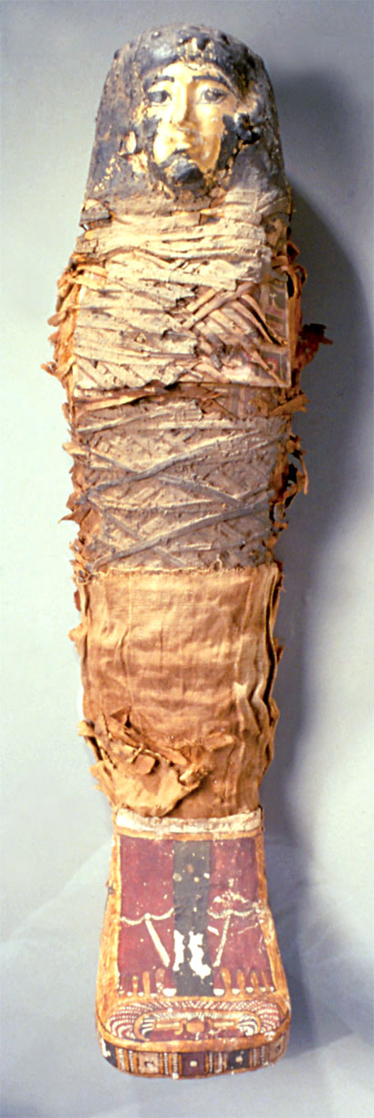
The findings from the British Museum mummy stoked Egyptologists' appetite to apply the same techniques to the mummy that has resided at the museum in San Jose for at least 75 years. The child is thought to have died between the ages of 4 and 6 and be from the Roman period, dating from the first century AD. This child was likely from a wealthy family, given the high-quality mummification and the gilded facemask.
Basic X-rays were taken of this mummy 20 years ago and revealed that the child had a hip abnormality. Newer imaging technologies, however, will provide much more information. In addition to helping to understand whether child burial techniques differed from those for adults—were kids buried, for instance, with different sorts of objects?—the modern techniques may help scientists to recreate how the child looked when alive.
Bioengineering professors may model how the child would have walked. And researchers at the Biocomputation Center may use the scans to do a facial reconstruction. "It is fantastic that we can provide the data for a kind of virtual autopsy," said Fahrig. "Using the latest medical technology, we can help curators examine the ancient world without harming the mummy."
The scans will be done Friday, but the results will not be immediately available. Scientists said that it could take months to compile and analyze the data.
Share This Story